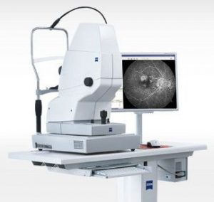
Corneal Mapping
Corneal topography is a painless, non-invasive medical imaging technique that maps the surface curvature of the cornea. The cornea is the clear front window of the eye, and its topography is critical in determining the quality of vision.
Corneal topography uses a computer-assisted device to create a three-dimensional map of the cornea. This map can be used to diagnose and treat various eye conditions, including astigmatism, keratoconus, and corneal scarring.
The procedure for corneal topography is quick and easy. The patient simply sits in front of a computer-assisted device and stares at a target. The device then projects a series of light patterns onto the cornea, which are reflected back to the device. The device uses these reflected patterns to create a three-dimensional map of the cornea.
Digital Retinal Imaging
Digital retinal imaging is a cutting-edge technology that uses a high-resolution camera to take pictures of the retina (the back of your eye). This is a painless and non-invasive procedure that can help your eye doctor detect eye diseases early, when they are most treatable.
The images taken during digital retinal imaging are stored electronically, which gives your optometrist a permanent record of the condition of your retina. This is important because many eye diseases, such as glaucoma, diabetic retinopathy, and macular degeneration, are diagnosed by detecting changes over time.
The advantages of digital retinal imaging include:
- It is quick, safe, and painless.
- It provides detailed images of the retina and sub-surface of your eyes.
- It provides instant, direct imaging of the form and structure of eye tissue.
- The image resolution is extremely high quality.
- It uses eye-safe near-infrared light.
- No patient prep is required.
Why is Digital Retinal Imaging Important?
Digital retinal imaging is important because it can help your eye doctor detect eye diseases early, allowing them to prescribe timely medications or laser treatment to prevent vision loss.
Digital retinal imaging can also be used to track the progression of eye diseases over time. This information can help your eye doctor make treatment decisions and monitor your overall eye health.
Cirrus Optical Coherence Tomography (OCT)
Cirrus OCT is a revolutionary imaging technology that uses light instead of sound waves to create detailed, high-resolution images of the back of the eye. This allows eye doctors to diagnose and monitor retinal disease more accurately than ever before.
This technology is particularly useful for the early detection of glaucoma, macular degeneration, and diabetic retinopathy. These conditions can cause irreversible damage to the eye, so early diagnosis is essential.
Cirrus OCT can also be used to monitor the effectiveness of treatment. By tracking changes in the retina over time, optometrists can make sure that treatment is working and adjust it as needed.
This is a painless, noninvasive test that takes about 10 minutes to perform. It is a safe and effective way to assess the health of your eyes.
If you are concerned about your eye health, talk to your optometrist about Cirrus OCT. This cutting-edge technology can help you protect your vision for a lifetime.
These are some of the benefits of Cirrus OCT:
- It can provide detailed images of the retina and optic nerve, which can help eye doctors diagnose and monitor eye diseases.
- It can be used to measure the thickness of the retinal nerve fiber layer (RNFL), which is a key indicator of glaucoma.
- It can be used to assess the structure and function of the macula, which is the part of the retina responsible for central vision.
- It can be used to track changes in the eye over time, which can help eye doctors assess the effectiveness of treatment.

Humphrey Visual Field Testing
A visual field test is a simple and painless way to measure the range of your peripheral or “side” vision. It can help to detect blind spots (scotomas), peripheral vision loss, and other visual field abnormalities.
The test is typically done in two parts. First, your eye doctor will perform a simple screening test. You will be asked to keep your gaze fixed on a central object while your eye doctor covers one eye. Then, you will be asked to describe what you see at the periphery of your field of view.
If the screening test indicates that you may have a visual field abnormality, your eye doctor may recommend a more comprehensive test using special equipment. This test involves placing your chin on a chin rest and looking ahead. Lights will be flashed on, and you will be asked to press a button whenever you see the light. The lights will be bright or dim at different stages of the test.
Each eye is tested separately, and the entire test takes about 15–45 minutes. The results of the test are used to create a computerized map of your visual field. This map can help your eye doctor to identify any areas of your vision that are deficient.

Zeiss Diagnostic Slit Lamp Examination with Anterior Segment Cameras
The ZEISS SL 220 slit lamp is a versatile and reliable tool for eye examinations. It features a popular tower design with LED illumination, providing years of reliable use. The SL 220 delivers superb optical and mechanical quality, as well as convenient operation and detailed, contrast-rich images. Wide-ranging accessories allow for individual setup to suit your preferences.
The SL 220 also features the ZEISS SL Imaging Module, which allows you to document any slit lamp examination with high-resolution images and videos. This is perfect for review, follow-ups, and patient education. The SL cam 5.0 camera and SL imaging software combine forces to deliver high-quality images and videos that are easy to share.

INMODE Lumecca Intense Pulsed Light treatment (IPL)
IPL is a safe and effective treatment for ocular rosacea, meibomian gland dysfunction, and inflammatory dry eye. It works by targeting the inflammatory blood vessels on the eyelid margin, which helps to reduce inflammation and improve tear production. IPL has also been shown to decrease demodex (a type of microscopic mite that naturally lives on human skin and hair follicles) and bacteria around the eyelids, which can further contribute to dry eye symptoms.
The IPL treatment is performed in an eye doctor’s office and is typically painless. You may experience some redness and swelling for a few hours after the treatment, but these side effects typically subside quickly.

INMODE Forma Radiofrequency treatment (RF)
RF is a treatment that uses radiofrequency energy to stimulate collagen production. This can help to improve the appearance of wrinkles and dark spots around the eyes. RF is also often used in conjunction with IPL to improve the overall results of the treatment.
The RF treatment is also performed in an eye doctor’s office and is typically painless. You may experience some redness and swelling for a few hours after the treatment, but these side effects typically subside quickly.
Combined IPL and RF Treatment
The combined IPL and RF treatment is a safe and effective way to improve the appearance of the eyes and reduce dry eye symptoms. The treatment is typically done in two stages, with the IPL treatment being performed first and the RF treatment being performed a few weeks later.
The combined treatment can help to:
- Reduce redness and inflammation
- Improve tear production
- Decrease demodex and bacteria around the eyelids
- Improve the appearance of wrinkles and dark spots
If you are considering IPL or RF treatment for your eyes, be sure to talk to your eye doctor about the best treatment option for you.

